DR. KIRAN KUMAR
MBBS, MD (General Medicine)
- Experience: 8 years
- Hospital: MEDI STAR Hospital
- Fee: ₹600
- Rating: ★ 4.0
Available today
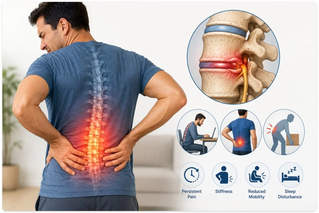
Short Description: Persistent pain or discomfort in the lumbar, thoracic, or cervical spine lasting for 12 weeks or longer, often stemming from spinal degeneration, muscle strain, or nerve compression.
Select the symptoms you are experiencing. We will route Normal Appointment, Urgent Request, or Emergency Guidance. This is navigation help, not a medical diagnosis.
Displacement or rupture of intervertebral disc gel pressing directly against adjacent spinal nerve roots in the lumbar region.
Age-related wear and breakdown of cartilage within spinal joints, causing friction, bone spurs, and chronic stiffness.
Progressive narrowing of the spinal canal that restricts space for the spinal cord and exiting spinal nerves.
Weak core abdominal/back muscles coupled with prolonged static sitting, placing elevated mechanical load on spinal structures.
Micro-tearing of supporting lumbar muscles and ligaments caused by heavy lifting, improper technique, or sudden twisting.
Evaluates posture, spinal range of motion, nerve reflexes, muscle strength, and sensation patterns to pinpoint nerve root compression or structural impairment.
High-detail soft-tissue imaging to detect herniated or bulging intervertebral discs, spinal canal stenosis, nerve root impingement, and ligament inflammation.
Standard radiographic imaging to assess bone alignment and identify vertebral fractures, joint narrowing, spondylolisthesis, or spinal osteoarthritis.
Cross-sectional bone imaging is used when MRI is contraindicated to closely examine complex bony structures, spinal canal dimensions, and facet joint arthrosis.
Diagnostic electrical tests to evaluate nerve function, measuring the speed and path of electrical impulses to confirm or rule out peripheral radiculopathy or neuropathy.
Lie flat on a firm mattress or floor with knees bent and supported by pillows, or sit in a supportive chair. Avoid complete, prolonged bed rest.
Apply a cold pack wrapped in a cloth or towel to the painful area for 15 to 20 minutes at a time, 3–4 times daily, to minimize localized inflammation and swelling.
After 48 hours, apply warm compresses or heating pads for 15–20 minutes to relieve muscular spasms. Perform gentle pelvic tilts or light walking as tolerated.
Take non-prescription pain relievers or anti-inflammatories (such as acetaminophen or ibuprofen) as directed on the label to manage acute discomfort.
Call emergency services immediately if the back pain follows high-impact trauma (e.g., car accident, fall) or is accompanied by sudden loss of bowel/bladder control or progressive leg paralysis.
Educational information only — not a personal prescription. Always consult a qualified clinician.
Targeted physical therapy focusing on core abdominal and spinal muscle strengthening, hamstring stretching, and postural retraining to support the spinal column.
Nonsteroidal anti-inflammatory drugs (NSAIDs) or muscle relaxants prescribed to reduce acute inflammation, ease localized muscle spasms, and control persistent spine pain.
Targeted fluoroscopy-guided corticosteroid injections into the spinal epidural space to relieve acute nerve root inflammation and radicular leg pain.
A minimally invasive procedure using heat to interrupt nerve conduction from painful facet joints, providing long-lasting pain relief.
Surgical procedures (such as microdiscectomy or spinal fusion) performed in severe cases to relieve nerve compression or stabilize unstable spinal segments.
Perform targeted core-strengthening and lumbar exercises (such as planks, pelvic tilts, and gentle spinal extensions) to build abdominal and trunk stability, reducing mechanical load on the spine.
Maintain an ergonomic workstation with adequate lumbar support and always lift heavy objects by bending at the knees and hips while keeping the back straight, avoiding sudden twisting movements.
Keep body mass within a healthy range to minimize continuous gravitational stress and mechanical pressure exerted on the lumbar spine, intervertebral discs, and surrounding joints.
tand up, stretch, and move around for 2 to 3 minutes every 30–45 minutes of continuous sitting to relieve sustained intra-discal pressure and prevent spinal muscle fatigue.
Avoid smoking, as nicotine restricts blood flow to intervertebral discs and accelerates spinal degeneration, and drink plenty of water to maintain disc hydration and elasticity.
Consume a diet rich in omega-3 fatty acids (salmon, walnuts), antioxidants (berries, leafy greens), and adequate calcium and vitamin D to support bone density and minimize systemic spinal inflammation.
Engage in regular low-impact physical activities like swimming, walking, or stationary cycling, paired with abdominal core-strengthening exercises (e.g., pelvic tilts, gentle planks) to stabilize the lumbar spine.
Utilize supportive seating with proper lumbar support, keep computer screens at eye level, maintain neutral spine alignment, and use a supportive mattress with a pillow under or between the knees during sleep.
Avoid sitting continuously for longer than 30–45 minutes, lifting heavy objects by bending at the waist, sudden twisting under load, high-impact jumping exercises, and smoking (which reduces disc hydration).
Take brief 2-minute standing or stretching breaks throughout the day, maintain a healthy body weight to lessen mechanical strain on spinal vertebrae, and practice stress-reduction techniques to alleviate muscle tension.
Back pain that persists continuously for over 6 to 12 weeks, worsens at rest, or wakes you up at night despite conservative physical therapy and medication.
Sharp, electric, or burning pain that shoots continuously down the back of the thigh and radiates below the knee into the foot or toes.
Sudden incontinence, loss of urinary/fecal control, or inability to urinate, indicating acute spinal cord or nerve root compression requiring emergency surgery
Severe loss of sensation or numbness localized to the groin, buttocks, inner thighs, or rectal area.
udden loss of muscle strength in the legs or inability to lift the front part of the foot while walking (foot drop), signaling severe nerve damage.
Based on the selected health concern, these specialists may be relevant. General Medicine is listed as a starting point for Chronic Back Pain — this is navigation help, not a diagnosis.
Care paths: Normal Appointment · Urgent Request · Emergency Guidance.
MBBS, MD (General Medicine)
Available today
MBBS, MD General Physician & Diabetologist
MBBS MD, Anaesthesia
MBBS, MS (Orthopedics), M.Ch
Senior consultant physiotherapist, HOD Physiotherapy and Rehabilitation
MBBS, MD (Internal Medicine)
Mumbai
Hyderabad
New Delhi
General Care
₹1,499
Executive
₹4,499
Saathora provides healthcare information to help you understand conditions and find care. It is not a substitute for professional medical advice, diagnosis, or treatment.
Completed appointment → Eligible for review. This becomes a Verified Patient Review for the doctor and hospital of this visit.
Accessibility details not listed yet.
Elderly priority assistance is available. Booked appointment times stay the same.
Saathora assistant